Equipment
CORE GIfMI offers cutting-edge MRI equipment backed by years of expertise and collaboration. From structural imaging to fMRI, MRS, MRE, and beyond - our versatile setup supports academic, clinical, and industry sponsored research across a wide range of applications.
Contact our Engineer
- Pieter Vandemaele
- +32 9 332 89 75
- pieter.vandemaele@ugent.be
Scanners

3T Siemens Prisma Fit MRI scanner
Our research-dedicated 3.0T Siemens Prisma Fit scanner is housed in the CORE GIfMI facility at Ghent University Hospital, Entry 55. The system is available 24/7 for your research needs.
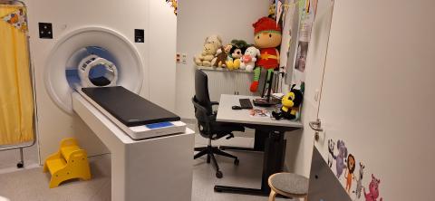
MRI Simulator - Free for Research
The MRI Simulator is available at no cost to all MRI researchers. It offers subjects a realistically preview of the scanning environment, helping improve comfort and compliance.
Psychophysiological Lab in the Magnet
CORE GIfMI is equipped with high-quality, dedicated equipment to support psychophysiological research within the MRI environment. This setup enables researchers to conduct advanced studies integrating neuroimaging with physiological measurements.
Visual System
- The BOLDScreen 32 UHD (Cambridge Research Systems Ltd.) is a high-performance, fully MR-compatible LCD display for research and participant entertainment.
- A 4x4 channel video switch enables seamless switching between multiple inputs and outputs - no cable changes needed.
Communication Systems
- The built-in communication system provides basic communication between participant and operator.
- The OptoActive II (Optoacoustics Ltc.) delivers high-fidelity audio with active noise cancellation (ANC) for EPI sequences.
- The FOMRI-III microphone ensures clear participant-operator communication, even during scans.
Response Devices
- Lumina 4G (Cedrus Corporation): Multiple 4-button layouts for response collection during fMRI, synchronized with MRI triggers.
- fMRI Trackball (Nata Technologies Inc.): enables cursor navigation during fMRI data acquisition - for tasks like rating scales or target pointing.
MRI Synchronization Triggering
- Custom-built Arduino-based hardware enables multi-channel signal conditioning and precise synchronisation between the scanner an external devices like the stimulus PC or BIOPAC system.
Eye Tracking
- The EyeLink 1000 Plus (SR Research Ltd.) is a video-based monocular eye tracker that delivers precise tracking inside the magnet when paired with the LCD video system.
Physiological Monitoring
- The built-in physiological monitoring system captures ECG and respiration signals for gating and physiological noise correction.
- The MP150 (Biopac Systems Inc.) with multiple modules supports simultaneous recording of various physiological, analog, and digital signals - fully synchronized with the MRI scanner for precise time labeling of the collected data.
- Available modules:
- MP150: the main module
- HLT100C: high-level output transducers interface
- STP100C: isolated digital interface
- UIM100C: universal interface module
- DA100C: general purpose transducer (for new RSP transducer)
- CO2100C: carbon dioxide measurement module
- RSP100C: respiration amplifier
- ECG100C-MRI: electrocardiogram amplifier
- PPG100C-MRI: photo plethysmogram amplifier
- EDA100C-MRI: electrodermal activity amplifier
Stimulus PC
- A high-performance PC with a dual-monitor setup gives you full control over your experiment. It can also play music or movies to keep participants relaxed during scans.
- Installed software:
- Matlab/Octave
- Conda (open-source package manager)
- PsychoPy (actively supported)
- Presentation (actively supported)
- Psychtoolbox (not actively supported)
- E-Prime (not supported)
- Other tools: on request
The Box
- Most of the peripheral equipment is connected to a custom-made rack-mount cabinet that provides the user with a single hardware interace for easy access and connectivity.
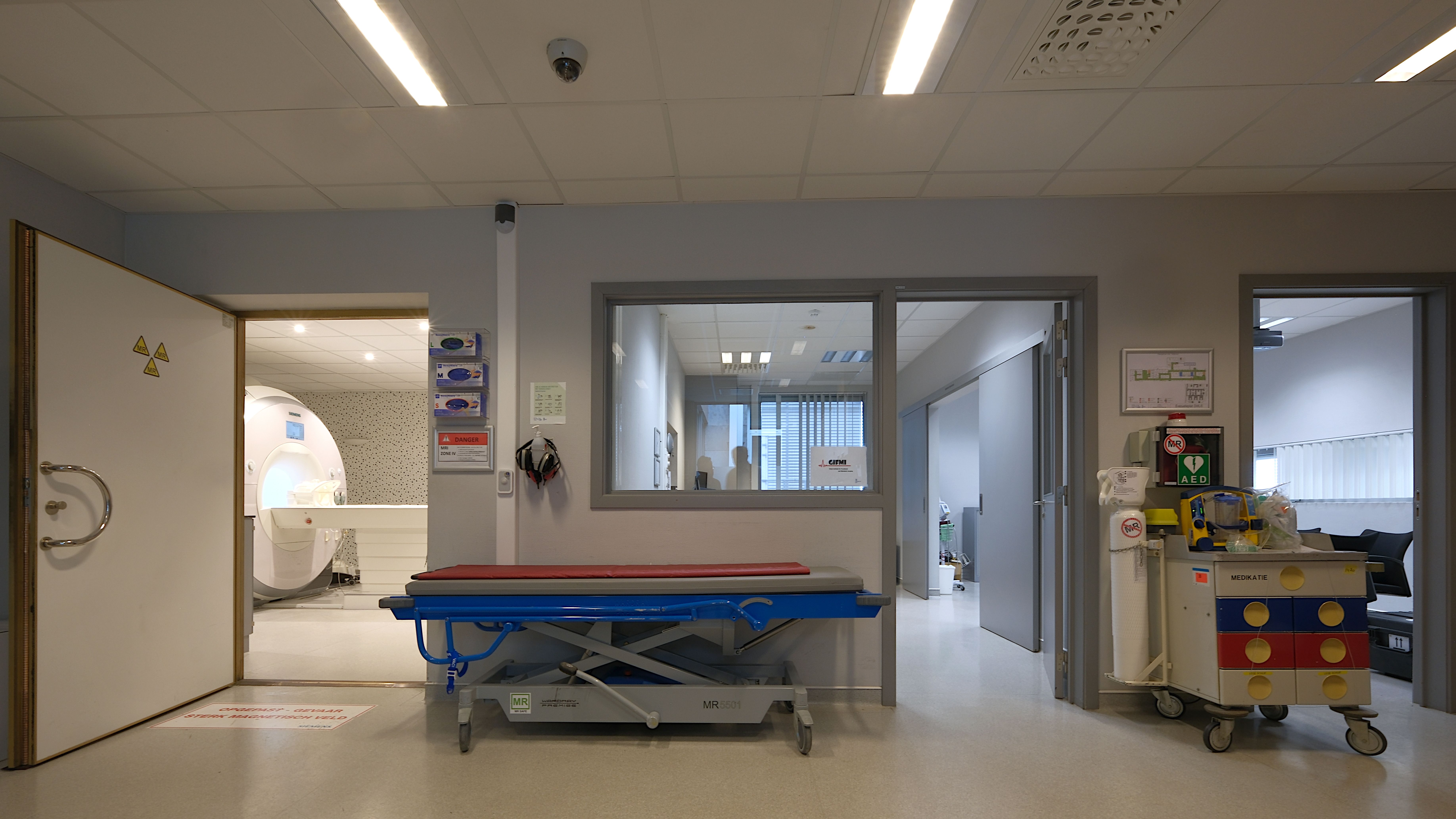
Facility Features
CORE GIfMI is housed in a dedicated building on the Ghent University Hospital campus. This enables researchers to fully focus on their tasks without disruption. Get familiar with our facility by visiting us virtually in the online tour.
Meeting Room Hippocampus
- Seats up to 12, equipped with a table and projector. Available by booking for researchers.
- Common uses:
- Participant briefing & debriefing
- Workspace for scanning buddies
- Meetings with CORE GIfMI Operational Team
- MRI Safety Training sessions
Control room
- The control room holds all MR control and advanced research equipment - fMRI, physiological monitoring, elastography, and more.
- A large window and video system provide constant visual monitoring of participants during MRI.
MRI Scanner Room
- Home to the MR scanner and peripheral equipment like video displays, audio systems, response systems, etc.
- Enclosed in a Faraday cage to block electromagnetic interference, ensuring optimal scanner performance.
- Audiovisual equipment can also be used to entertain participants when allowed.
TMS room
- The facility includes a Transcranial Magnetic Stimulator (TMS) managed and operated by the Department of Psychology.
Other equipment
CORE GIfMI also houses specific hardware and software for specialist MR data acquisition.
Magnetic Resonance Elastography
- Magnetic Resonance Elastography (MRE) is technique to quantify the stiffness of tissue in the body. The MR Elastography device (T.H.E.A-Devices GmbH) generates low-frequency vibrations in tissue. A special sequence and post-processing software calculates the elastogram. More to find on the Techniques page.
MRI Phantoms
- Small & Large ACR-NEMA for structural imaging
- FUNSTAR for fMRI
- QASPER (Quantitative Arterial Spin Labelling Perfusion Reference, Gold Standard Phantoms Ltd) is a calibration and quality assurance standard for MRI perfusion measurements using Arterial Spin Labelling (ASL). It simulates the process of delivery of arterial blood to an organ in a controlled and reproducible manner. More information can be found on the manufacturer's website.
Computing Facilities
CORE GIfMI does not provide computing facilities but collaborates closely with HPC-UGent, the High Performance Computing infrastructure of Ghent University. As a strategic partner of the Flemish Supercomputing Center, HPC-UGent offers researchers access to a variety of image processing tools on its HPC infrastructure.
Some tools available at the HPC-UGent are
- Neurodesk: a server-side containerized environment for neuroimaging data analysis, providing access to all major open-source neuroimaging tools.
- Major (neuro)imaging processing software like FSL, MRtrix, SPM, FreeSurfer, nipype, nilearn
- Preprocessing tools like ANTs, SimpleElastix
- Image format tools like pydicom, dicom2nifti, NiBabel, MedPy
- High-level programming languages like Python, Matlab, R, Lua, Go
- Tools can be added on request by the HPC-UGent team
Data Management
CORE GIfMI is currently working on a XNAT (Extensible Neuroimaging Archive Toolkit) instance to manage all MR research images and metadata.
Once fully operational, this system will provide researchers with a structured and secure platform for storing, sharing, and processing imaging data.